
Research Overview
MD Biophysics is grounded in research at the intersection of photomedicine and cellular biology.
Where it startedThe Science of Light
In the 1970s, biophysicist Fritz-Albert Popp discovered that cells emit coherent light, known as biophotons.
“We now know, for example, that quanta of light can initiate, or arrest, cascade-like reactions in the cells, and that genetic cellular damage can be virtually repaired, within hours, by faint beams of light. We are still on the threshold of fully understanding the complex relationship between light and life, but we can now say emphatically, that the function of our entire metabolism is dependent on light.”
— Dr. Fritz-Albert Popp
Featured Research
MD Biophysics Photobiomodulation Plasma (PPT)/Very Small Embryonic like (VSEL) Antibody Marker Trend Analysis
Dawn DeSylvia, MD¹ and Ian Mitchell²
¹The Center for Whole Health, Agoura Hills, CA
²Wizard Sciences, Bartlesville, OK
Corresponding Author Contact Information: Dr Dawn DeSylvia, drdesylvia@wholelifehealthmd.com
Abstract
Background: Photobiomodulation (PBM) therapy has demonstrated therapeutic potential in promoting cellular repair, modulating inflammation, and enhancing mitochondrial function. Platelet-rich plasma (PRP) is widely used in regenerative medicine due to its concentration of growth factors and cytokines. Very small embryonic-like stem cells (VSELs), a rare population of pluripotent stem cells present in adult tissues, have emerged as a potential contributor to tissue regeneration. While PBM and PRP are used in combination, how VSELs or Multi-lineage stress enduring (MUSE) cells are at play, and the biological mechanisms underlying their synergistic effects remain incompletely characterized.
Objective: This exploratory pilot study aimed to evaluate whether application of the MD Biophysics laser to autologous PRP is associated with measurable changes in VSEL-related antibody marker expression, and to identify directional trends to inform future controlled studies.
Methods: PRP samples were collected from participants across seven test dates (July 2024 to February 2025), yielding 18 participant-session datasets. Samples were analyzed before (Pre) and after (Post) laser application using flow cytometry conducted at a UCLA Flow Cytometry Laboratory. Four VSEL-associated antibody markers were assessed: CD45⁻CD34⁺, CXCR4⁺, CD133⁺, and SSEA-4⁺. Analyses were descriptive and focused on paired differences and directional trends due to the exploratory design and absence of a control group.
Results: Three of four VSEL-associated markers (CXCR4⁺, CD133⁺, and SSEA-4⁺) demonstrated a group-level increase in median paired differences following laser application. Directional increases were observed in 12/18 sessions for CXCR4⁺, 10/18 for CD133⁺, and 9/18 for SSEA-4⁺. CD45⁻CD34⁺ showed a near-equal distribution of increases and decreases. Ki-67 positivity indicated the presence of viable, proliferative cells. While no findings reached statistical significance due to limited sample size, consistent directional trends were observed across multiple markers.
Conclusion: Application of PBM to autologous PRP was associated with directional increases in multiple VSEL-associated antibody markers, suggesting a potential role for stem cell activation or mobilization in the mechanism of action. Although preliminary and not statistically powered, these findings provide hypothesis-generating evidence supporting further investigation. The observed trends informed iterative protocol refinement and establish a foundation for future controlled, adequately powered studies to evaluate clinical efficacy and underlying biological mechanisms.
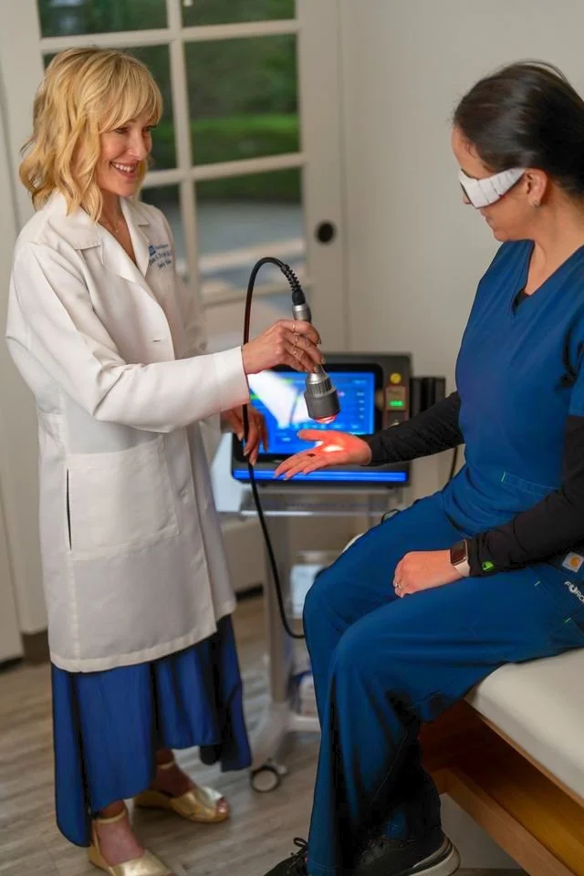
Real results
Before
Long standing psoriasis before treatment
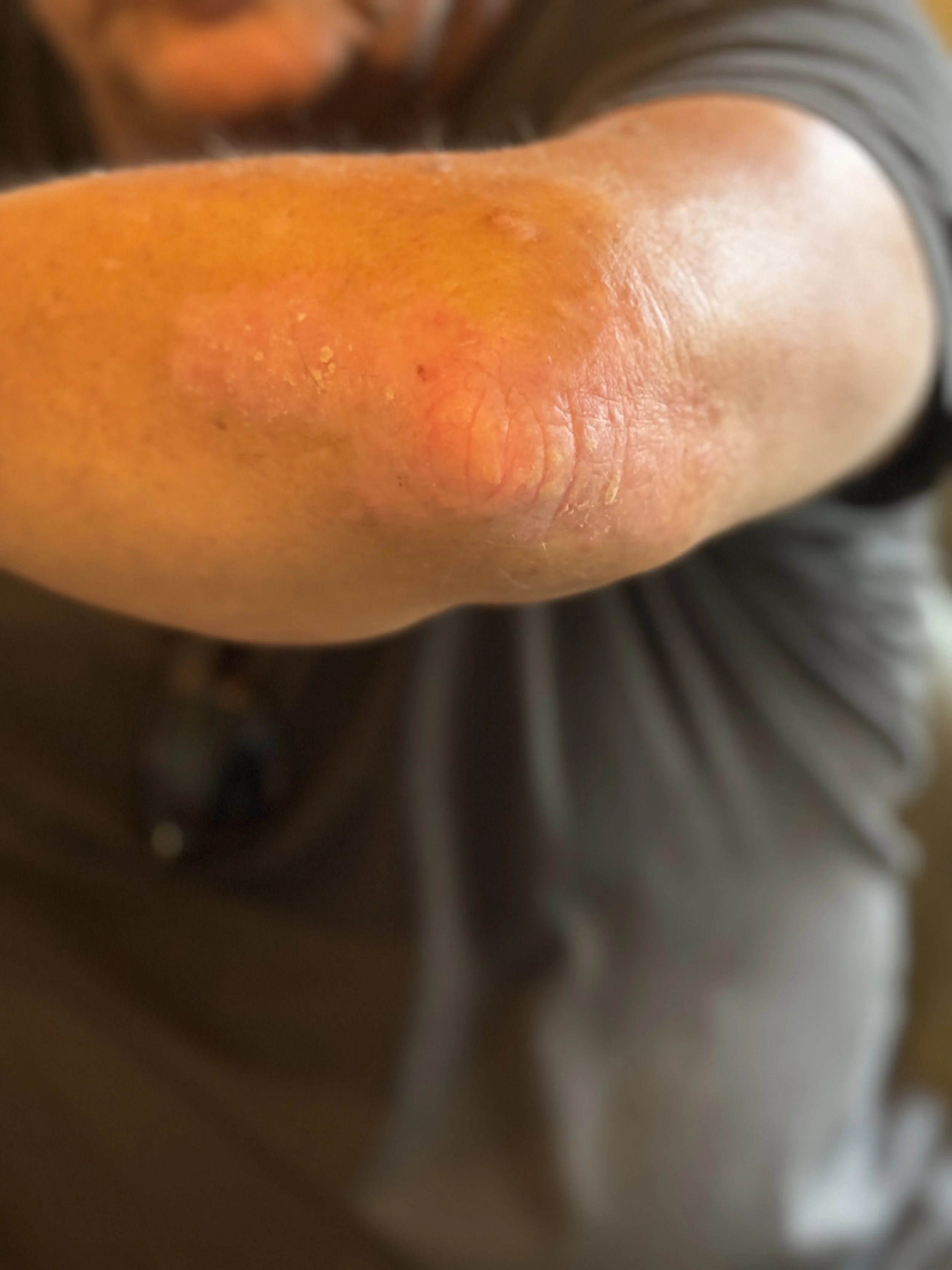
After
One day after single treatment
“In the quiet language of cells, the body remembers its wholeness, and begins again.”